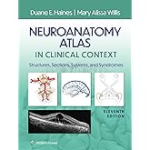
Bought this for my med school course in neuroscience and behaviour. The quality of the images and information is excellent. The best feature by far in my opinion is that the authors have sections dedicated to lesions of specific areas that show you visually which tracts/nuclei can be affected and why that would produce specific symptoms. Great resource for doing problem-based or case-based learning exercises and for Step 1 prep. There are USMLE-style review questions in the book and more available online using the scratch off code inside the cover.

Download the free Kindle app and start reading Kindle books instantly on your smartphone, tablet or computer—no Kindle device required.
Read instantly on your browser with Kindle for Web.
Using your mobile phone camera, scan the code below and download the Kindle app.

Follow the author
Something went wrong. Please try your request again later.
OK
Neuroanatomy in Clinical Context: An Atlas of Structures, Sections, Systems, and Syndromes Paperback – 19 May 2014
by
Haines
(Author)
Sorry, there was a problem loading this page.Try again.
Neuroanatomy in Clinical Context, Ninth Edition provides everything the student needs to master the anatomy of the central nervous system, all in a clinical setting. Clear explanations; abundant MRI, CT, MRA, and MRV images; full-color photographs and illustrations; hundreds of review questions; and supplemental online resources combine to provide a sound anatomical base for integrating neurobiological and clinical concepts. In thus applying neuroanatomy clinically, the atlas ensures student preparedness for exams and for rotations. This authoritative approach�combined with such salutary features as full-color stained sections, extensive cranial nerve cross-referencing, and systems neurobiology coverage�sustains the legacy of this revolutionary teaching and learning tool as the neuroanatomy atlas. New and hallmark features elucidate neuroanatomy and systems neurobiology for course success! NEW! Chapter on Herniation Syndromes decodes the elegant relationship between brain injury and resulting deficit. NEW! Clinical information integrated throughout the text is screened in blue for quick identification on the page. NEW! Enhanced clinical images emphasize clarity and detail like never before, including full-color images replacing many in black and white, higher-resolution brain scans, and reprocessed spinal cord and brainstem images. MRIs complement full-color anatomical illustrations, allowing for visualization of structures both as they appear to the unaided eye and on imaging studies. Unique, full-color illustrations integrate clinical images of representative lesions with the corresponding deficits highlighted. Full-color stained sections facilitate the easy identification of anatomical features. Dozens of pathway drawings superimposed over MRIs connect structure with function of neural pathways. Located on thePoint, this atlas�s companion website offers a variety of supplemental learning resources to maximize study and review time! Question bank featuring over 280 USMLE-style and chapter-review style questions Bonus dissection photographs and brain slice series
- ISBN-101451186258
- ISBN-13978-1451186253
- Edition9th
- PublisherLippincott Williams & Wilkins USA
- Publication date19 May 2014
- LanguageEnglish
- Dimensions22.86 x 1.91 x 31.12 cm
- Print length368 pages
Customers who viewed this item also viewed
Page 1 of 1 Start againPage 1 of 1
Product details
- Publisher : Lippincott Williams & Wilkins USA; 9th edition (19 May 2014)
- Language : English
- Paperback : 368 pages
- ISBN-10 : 1451186258
- ISBN-13 : 978-1451186253
- Dimensions : 22.86 x 1.91 x 31.12 cm
- Best Sellers Rank: 1,065,178 in Books (See Top 100 in Books)
- 315 in Neuroscience Textbooks
- 405 in Anatomy Textbooks
- 1,269 in Medical Anatomy
- Customer Reviews:
About the author
Follow authors to get new release updates, plus improved recommendations.

Discover more of the author’s books, see similar authors, read book recommendations and more.
Customer reviews
4.6 out of 5 stars
4.6 out of 5
129 global ratings
How are ratings calculated?
To calculate the overall star rating and percentage breakdown by star, we don’t use a simple average. Instead, our system considers things like how recent a review is and if the reviewer bought the item on Amazon. It also analyses reviews to verify trustworthiness.
Top reviews from Australia
There are 0 reviews and 0 ratings from Australia
Top reviews from other countries
-
 MarkoReviewed in Germany on 21 May 2018
3.0 out of 5 stars Great but wasn’t new, damaged item
Verified PurchaseFantastic book, no doubt about it. The reason for 3 stars is due to the fact that the book that arrived was damaged and clearly not new, very disappointed
 KQReviewed in Canada on 13 July 2016
5.0 out of 5 stars Great supplement for medical school
Verified PurchaseThe best neuroanatomy textbook out there. Great for medical school neuroscience, shows relevant CT's and MRIs with great explanations. The explanations for the clinical syndromes are also very thorough.


